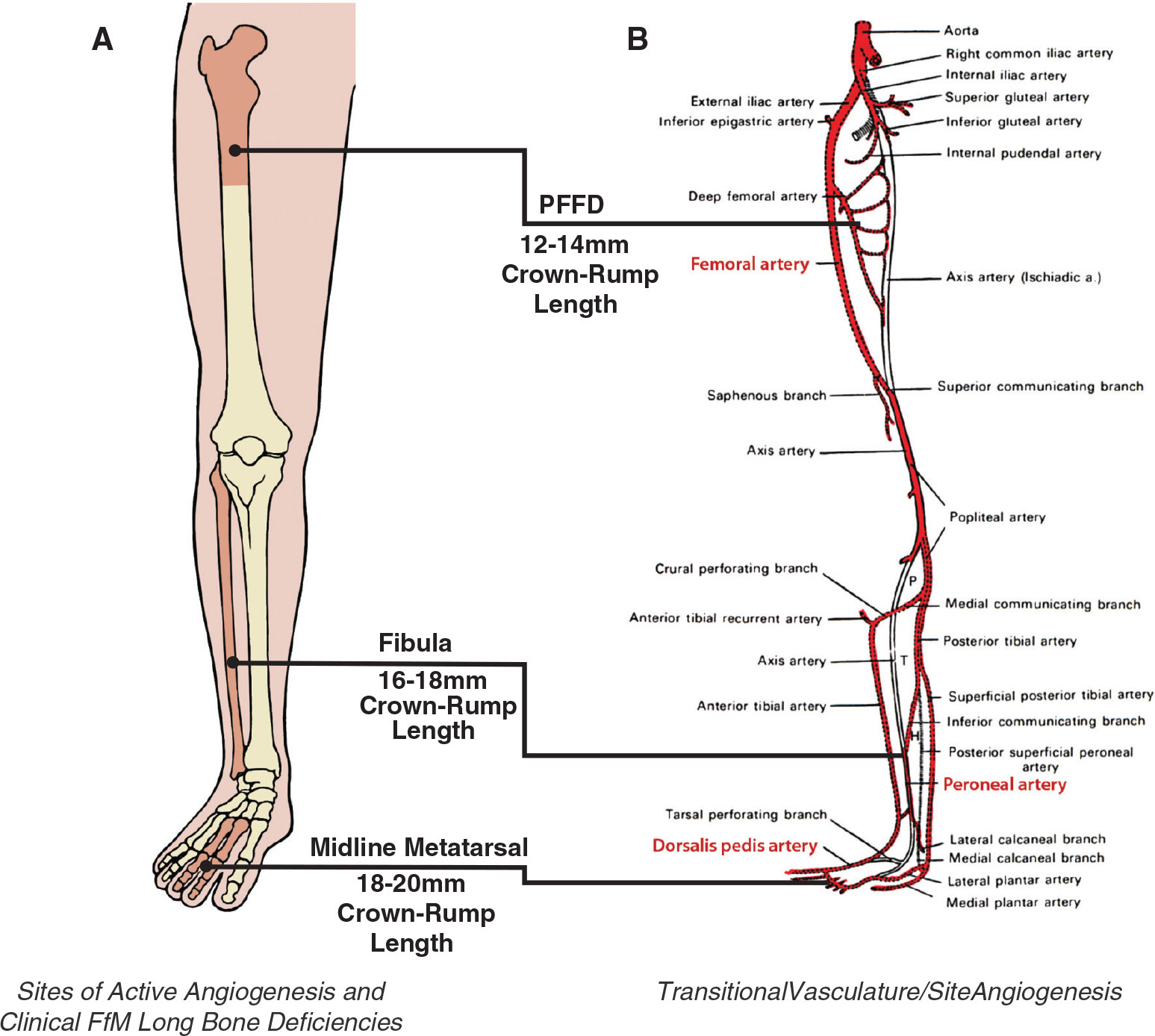
OrthoBuzz occasionally receives news and perspectives from members of the orthopaedic community on topics of interest to the JBJS readership. Orthopaedic surgeon Dr. David R. Hootnick of SUNY Upstate Medical University in Syracuse, New York, is a co-author of a recent review article in the journal Birth Defects Research that examines the origins of congenital long-bone deficiencies in the lower-limb. He and his co-authors emphasize embryonic arterial maldevelopment—injury to the developing blood vessels—as the root cause leading downstream to a spectrum of skeletal deficiencies. Dr. Hootnick shared more on this with OrthoBuzz. His comments have been edited and condensed.
What inspired your investigative work in this area?
My fellowship at the Great Ormond Street Hospital for Sick Children in London in 1975 centered on a study of children with congenital short limbs and fibular deficiency1. A number of the patients had obvious radiographic deficiencies of the second and third metatarsals2. Those missing metatarsals being midline rather than lateral conflicted with the standard literature on the classification of “fibular hemimelia”3,4. This was an “aha” moment for me.
It was my very good fortune the following fellowship year to return to Boston, to the New England Baptist Hospital. Amazingly, I learned that, among limbs of children with severe proximal femoral focal deficiency [PFFD]—a recently named entity—as many as 80% displayed the fibular deficiency that I had studied in London. That didn’t make any sense in terms of our understanding of how limbs are formed. The embryology of the development of the blood vessels in Gray’s Anatomy from a century ago did correspond syndromically to the 3 regions of long-bone radiographic deficiency5.
In a recent review article in Birth Defects Research, Embryonic Vascular Dysgenesis: The Origin of Proximal Femoral Focal Deficiency, you and your co-authors explore embryonic vascular dysgenesis as a root cause of a spectrum of congenital long-bone deficiencies. You write that PFFD “is the most proximal manifestation of a syndrome involving Congenitally Shortened lower Limbs (CSL), which also affects the fibula and midline metatarsals. This pattern of congenital human long bone deficiencies corresponds, in a time dependent manner, to the failed ingrowth pathways of new blood vessels of the growing embryonic limb”6.
Jonathan Cohen and Henry Cowell, then Pediatrics editors with JBJS, helped us with publication of a paper fundamentally related to this topic in 1980, Vascular Dysgenesis Associated with Skeletal Dysplasia of the Lower Limb [showing an abnormal arterial pattern in 3 patients with fibular deficiencies when evaluated angiographically]7. A more recent paper regarding the variable fibular deficiencies was published in 2020 in The Anatomical Record, Congenital Fibular Dystrophisms Conform to Embryonic Arterial Dysgenesis8.
The bottom line is that the foundations of all of the long bones are present before the embryonic limb grows out. The femur, fibula, and metatarsals then develop in conformity with their available blood supplies. Conversely, the models of the long bones are present before the limb grows out and can develop only after being supplied by their blood vessels. Congenital malformations of the femur, fibula, and metatarsals all follow the dystrophic pathways created by their remnant “trace fossil” arteries.
What is a key point you wish to emphasize for the orthopaedic readership?
Those who attempt to lengthen congenitally shortened limbs must be aware of the biologic limitations of surgery due to the missing blood vessels.
References
- Hootnick D, Boyd NA, Fixsen JA, Lloyd-Roberts GC. The natural history and management of congenital short tibia with dysplasia or absence of the fibula. J Bone Joint Surg Br. 1977 Aug;59(3):267-71.
- Hootnick DR, Levinsohn EM, Packard DS Jr. Midline metatarsal dysplasia associated with absent fibula. Clin Orthop Relat Res. 1980 Jul-Aug;(150):203-6.
- Frantz CH, O’Rahilly R. Congenital Skeletal Limb Deficiencies. J Bone Joint Surg Am. 1961 Dec;43(8):1202-24.
- Reyes BA, Birch JG, Hootnick DR, Cherkashin AM, Samchukov ML. The Nature of Foot Ray Deficiency in Congenital Fibular Deficiency. J Pediatr Orthop. 2017 Jul/Aug;37(5):332-7.
- Hootnick DR, Vargesson N. The syndrome of proximal femur, fibula, and midline metatarsal long bone deficiencies. Birth Defects Res. 2018 Sep 1;110(15):1188-93.
- Hootnick DR, Vargesson N, Horton JA, Chomiak J. Embryonic Vascular Dysgenesis: The Origin of Proximal Femoral Focal Deficiency. Birth Defects Res. 2025 Apr;117(4):e2465.
- Hootnick DR, Levinsohn EM, Randall PA, Packard DS Jr. Vascular dysgenesis associated with skeletal dysplasia of the lower limb. J Bone Joint Surg Am. 1980 Oct;62(7):1123-9.
- Hootnick DR. Congenital fibular dystrophisms conform to embryonic arterial dysgenesis. Anat Rec (Hoboken). 2020 Nov;303(11):2792-2800.
Image reproduced from: Horton JA, Krisch MF, Levinsohn EM, Hootnick DR. Lateral Epiphyseal Narrowings with Absent Fibula Conform to a Vascular Pattern Deficiency. JPOSNA. 2022 Aug 1; 4(3): 515. © 2022 JPOSNA. Published by Elsevier Inc. This is an open access article distributed under the Creative Commons Attribution License: https://creativecommons.org/licenses/by-nc-nd/4.0/


