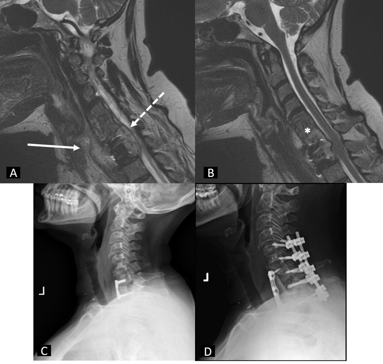
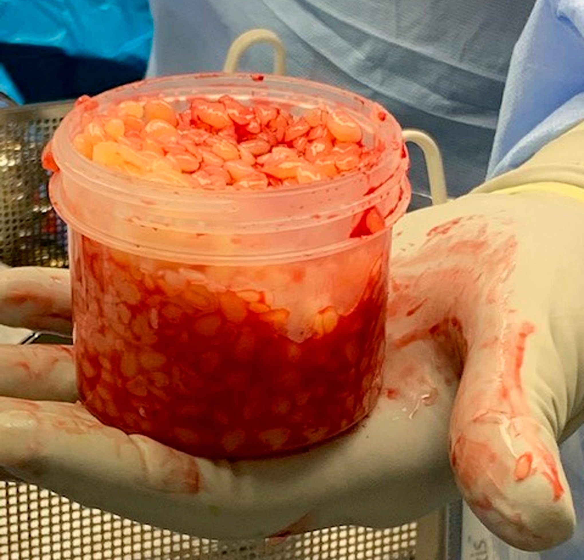
Recent reports in JBJS Case Connector describe 2 orthopaedic cases involving the formation of “rice bodies,” fibrinous loose bodies with a rice-like appearance. As discussed
Category: JBJS CC

JBJS Case Connector is looking to expand its roster of reviewers with expertise in the pediatrics, spine, hand & wrist, and infection subspecialties. Case Connector,

In this post, Dr. Thomas Bauer, Co-Editor of JBJS Case Connector, discusses a recent report of 2 cases of Mycobacterium tuberculosis osteomyelitis after spinal fusion

