JBJS Deputy Editor for Social Media Dr. Matt Schmitz discusses a new study assessing the use of post-reduction MRI measurements to help predict residual acetabular dysplasia in infants with developmental dysplasia of the hip.
Developmental dysplasia of the hip (DDH) is a continuum that includes hips that are located at birth but dysplastic, through hips that are dislocated at birth and won’t reduce with bracing early in life. Patients may require treatment in the operating room with either closed reduction and casting or open reduction (to remove barriers to reduction) and casting. In the past, we commonly used computed tomography (CT) to check the reduction, but that has been replaced over the past decade-plus with post-reduction magnetic resonance imaging (MRI).
One of the questions that remains in the treatment of DDH is which children with successful open or closed reduction are going to have residual acetabular dysplasia (RAD) needing further surgical treatment down the road?
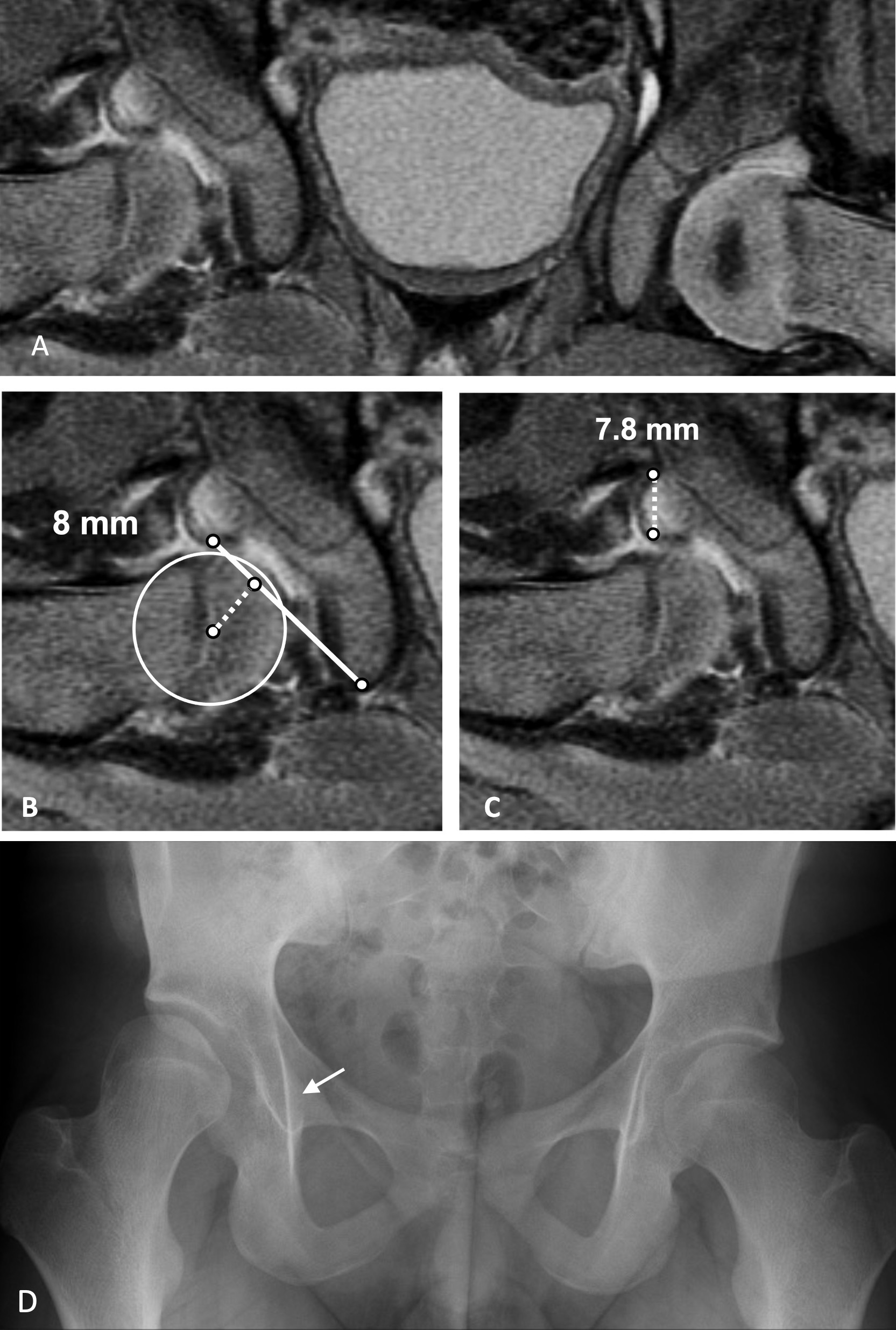
In the January 17, 2024 issue of JBJS, Schmaranzer et al. report the results of a retrospective study at Boston Children’s Hospital in which they examined whether hip concentricity and acetabular morphological measurements on post-reduction MRI are associated with RAD at a minimum of 10 years of follow up. The article is available at JBJS.org:
After exclusions and loss to follow-up, the authors reviewed 47 hips in 42 patients with a mean follow-up of 12 years. Twenty-nine (62%) of the 47 hips underwent closed reduction, and 18 (38%) had open reduction. The median patient age at reduction was 6.3 months; 83% of the hips were in female patients.
RAD was defined as additional surgery for residual dysplasia within the follow-up period or a Severin grade1 of >2. Patients with a Severin grade of ≤2 and no subsequent surgeries were allocated to the “no RAD” group.
The authors found that 24/47 (51%) had no RAD, while 23/47 (49%) developed RAD at a median of 11.4 years. Five of the 23 hips with RAD met the Severin grade criterion, while 18/23 (78%) had undergone surgery, with RAD confirmed by radiographic measurements prior to surgery. There was a significant difference in the median age between the groups, with the patients in the RAD group being older than those in the no-RAD group (7.3 vs 3.6 months; p = 0.009)
Most of the parameters from the coronal and axial MRI scans differed between the hips that ended up developing RAD and those that did not. A receiver operating characteristic (ROC) curve analysis was used to quantify the ability of the measurements to distinguish between patients with and without RAD, and the area under the ROC curve (AUC) was used to assess the diagnostic accuracy of measurements in predicting RAD. Cutoff values providing the best discrimination were calculated using the Youden index.
The highest discriminatory ability was found for the femoroacetabular distance in the coronal plane (AUC, 0.95; 95% CI, 0.90 to 1.00) with a 5-mm cutoff, and limbus thickness (AUC, 0.91; 95% CI, 0.83 to 0.99) with a 4-mm cutoff. Femoroacetabular distance in the coronal plane represents displacement from a perfectly concentric reduction (due to dysplasia on the acetabular side at the time of reduction), while the limbus thickness may show the degree of dysplasia and could lead to less remodeling potential.
The study highlights that a good concentric reduction (as demonstrated by the femoroacetabular distance in the coronal plane) is paramount and can help with continued development of the femoroacetabular joint. The authors acknowledge that these detailed measurements may be difficult to replicate clinically, but as imaging techniques and measuring ability improve, this could lead to new measures in predicting the need for future surgical procedures. Gone are the days where we rely on an arthrogram and medial dye space to assess the adequacy of our reduction in DDH on a 2-dimensional image. Now we have 3-dimensional advanced imaging and advanced measurements to help potentially guide our counseling of the families of these young patients.
Emmanouil Grigoriou, MD, and William J. Shaughnessy, MD share additional thoughts on this study in their commentary article, What Is the Relevance of Post-Reduction MRI Today and Where Do We Go from Here?
Read the study by Schmaranzer et al.: Hip Morphology on Post-Reduction MRI Predicts Residual Dysplasia 10 Years After Open or Closed Reduction
JBJS Deputy Editor for Social Media
References
- Severin E. Congenital dislocation of the hip; development of the joint after closed reduction. J Bone Joint Surg Am. 1950 Jul;32-A(3):507-18.


