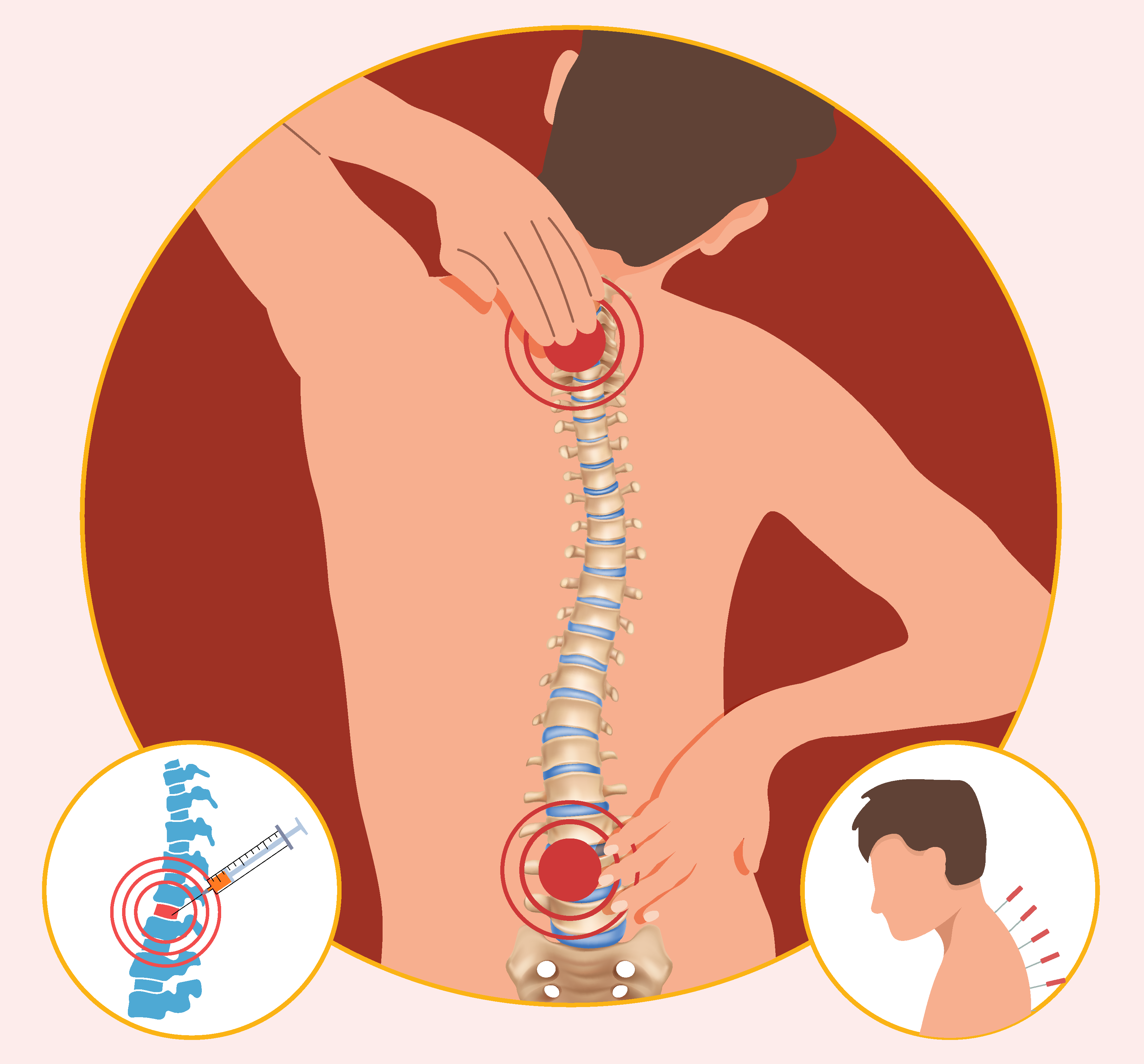
This post spotlights 2 new studies in JBJS that examined outcomes of spine surgery using nationwide databases. Patients with degenerative lumbar disease may turn to
Category: Spine
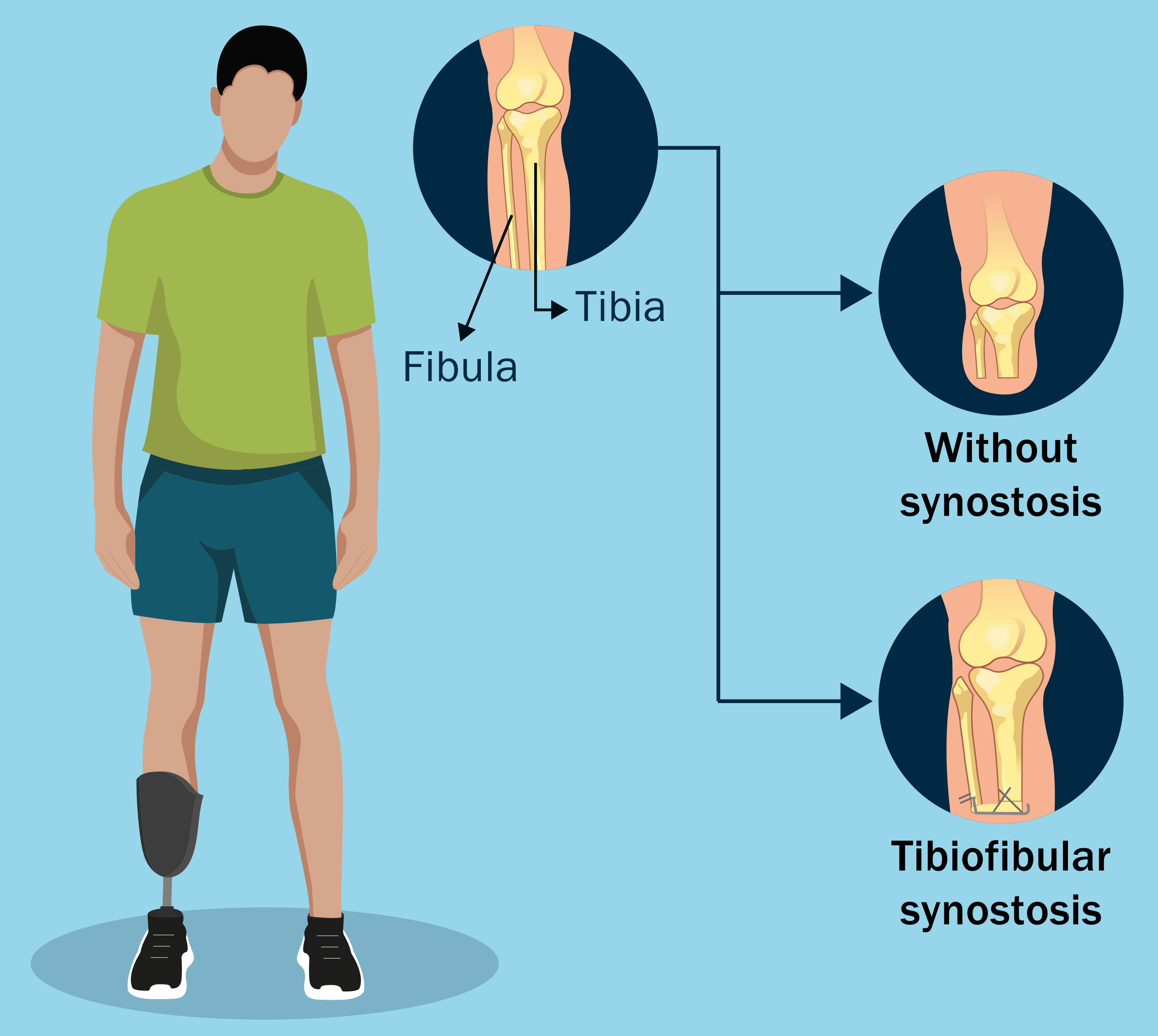
Dr. Matt Schmitz, JBJS Deputy Editor for Social Media, discusses 3 recent studies that reflect the rigorous standards, and challenges, of randomized clinical trials (RCTs).
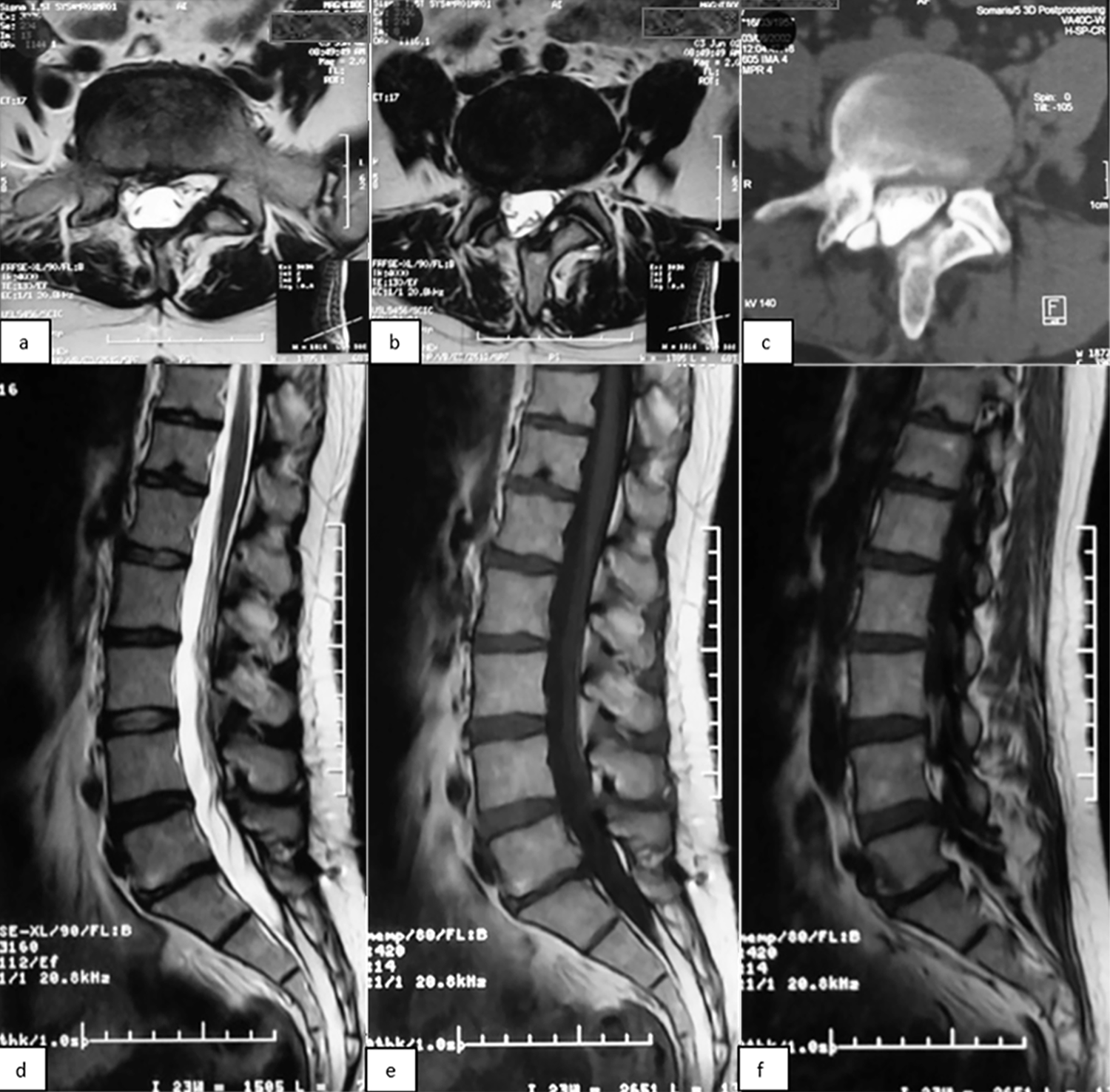
Investigators in France evaluated the long-term outcomes of 1- and 2-level total disc arthroplasty (TDA) in patients with chronic lumbar degenerative disc disease. A total
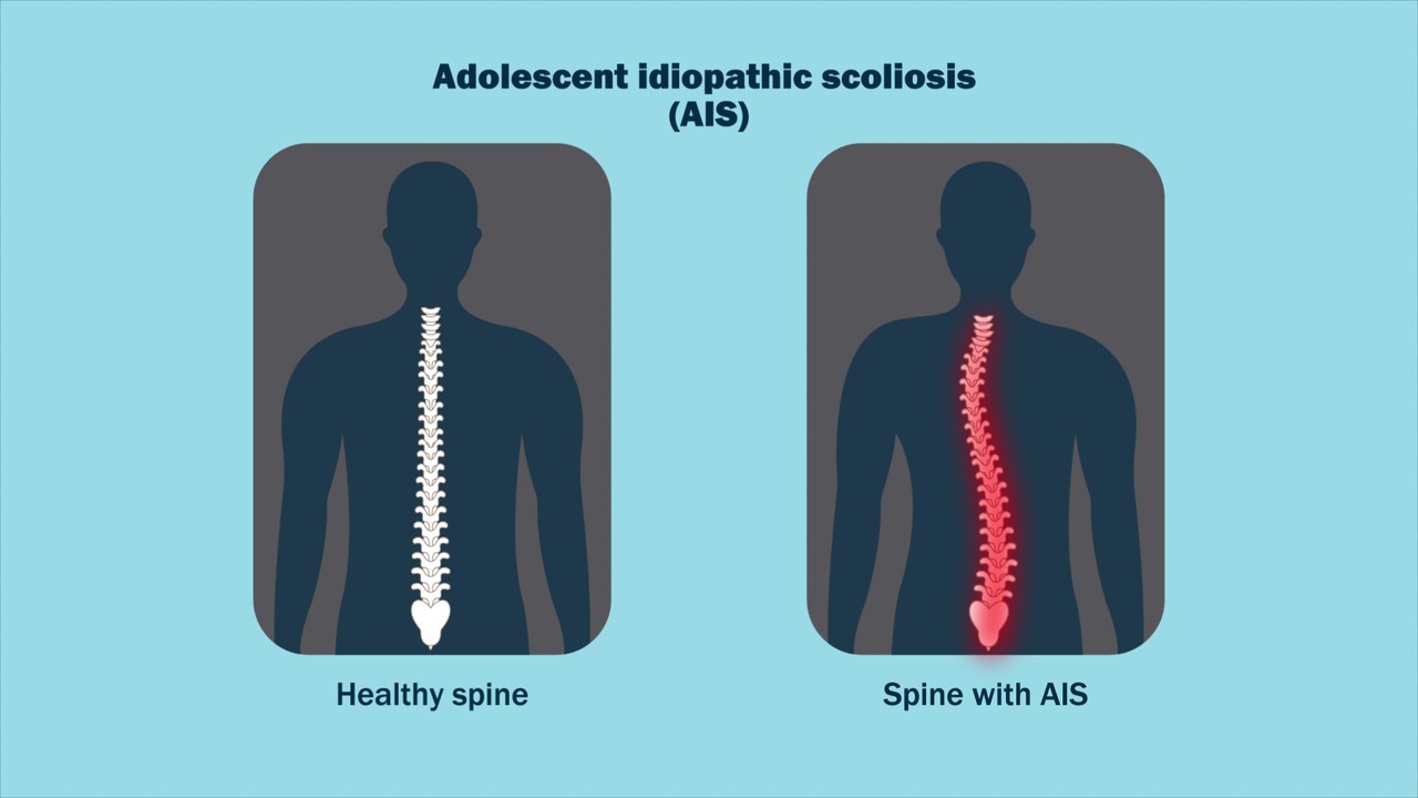
For patients with adolescent idiopathic scoliosis (AIS), could certain aspects of the spine examination be reliably conducted via telemedicine? Dr. Matt Schmitz, JBJS Deputy Editor
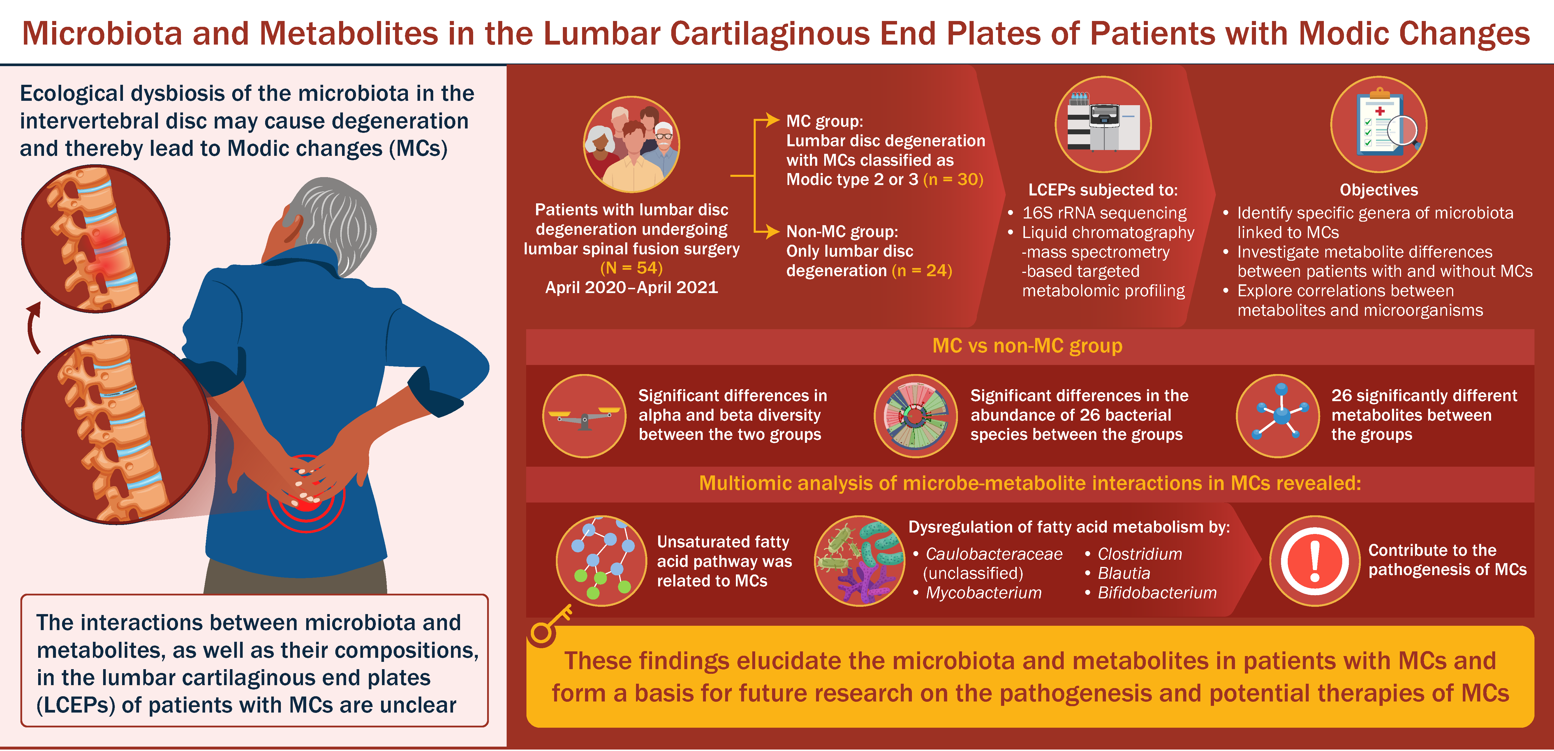
As reported recently on OrthoBuzz, a study by Nian et al. provides novel exploration of microbiota and metabolites in patients with Modic changes—changes in vertebral

